BackgroundThe onset of menarche is influenced by various factors, including genetic,
morphological and socioeconomic factors.
ObjectivesThe study aimed to examine the differences in adiposity levels and fat distribution
between early, average, and late maturing girls from Kolkata, India.
Sample and Methods936 Bengali girls included in a cross-sectional study were categorized as early,
average or late maturing. The examination was school-based and conducted from 2005 to
2011. Six skinfolds (biceps, triceps, subscapular, suprailiac, abdominal and calf) were
measured. Trunk-to-limbs, trunk-to-total, abdominal-to-trunk skinfold ratios, and total
adiposity were calculated. Statistical differences between all menarche categories were
assessed using Student t-test or Mann-Whitney test.
ResultsEarly maturing girls were characterized by greater overall (% BF: early=24.3;
average=24.0; late=23.8; p>0.05) and abdominal adiposity (as represented by skinfold
thicknesses and values of studied indicators), compared to those with late or average
age at menarche.
ConclusionsEarly menarche was associated with a tendency towards central adiposity and thus,
increased risk of abdominal obesity. Future research should explore the association
between the age at menarche and metabolic characteristics in ethnically diverse
populations. Longitudinal studies and studies conducted on large cohorts are
particularly valuable. It would be beneficial to adjust the results for factors such as
diet or physical activity, as well as for ethnic characteristics in relation to the
body's tissue composition.
Keywords: fat distribution, menarche, body fat, skinfold thickness
Conflicts of interest: There are
no conflicts of interest.
Citation: Kryst, Ł. et al. (2023). Adiposity level, fat distribution and age at menarche in Bengali girls from
Kolkata, India. Human Biology and Public Health 1. https://doi.org/10.52905/hbph2023.1.53.
Copyright: This is an open access article distributed under the terms of the Creative Commons Attribution License which permits unrestricted use, distribution, and reproduction in any medium, provided the original author and source are credited.
In the examined group of Indian girls, early menarche was associated with a tendency
towards central adiposity and an elevated probability of abdominal obesity. It is
important for future research to explore the association between age at menarche and
metabolic characteristics in ethnically and socioeconomically diverse populations.
Contents
Introduction
Menarche is influenced by a plethora of factors which can be divided into genetic,
morphological and environmental/socioeconomic aspects. For instance, it has been suggested
that up to 50–80 % of the variance in human pubertal timing is determined by genetic factors
(Gajdos et al. 2010). High level of genetic
control over the timing of menarche is also confirmed by its estimated heritability of
50% (Demerath et al.
2013).
Is has been suggested that genes analyzed as regulatory candidate for the age at menarche
(e.g., LIN28B, FTO, TNNI3K, GPRC5B) may also be linked to various anthropometric
characteristics such as body height and mass suggesting a common genetic basis for these
traits (Elks et al. 2010; Perry et al. 2014). Some alleles related to higher Body Mass Index
(BMI) and Waist-to-Hips Ratio (WHR) show at least nominal associations with an earlier onset
of menarche. This, in turn, suggests that menarche loci can generally have a pleiotropic
effect on growth (Elks et al. 2010).
It should also be stressed, that the described genetic factors closely interact with
environmental and socioeconomic aspects in regulating the timing of puberty (Gajdos et al. 2010). The acceleration of the biological
maturation appears to be highly correlated with the economic development of a country, and
consequently, the improvement of living conditions (Deardorff et al. 2014). Indian girls coming from more affluent families tend to
experience menarche at an earlier age, compared to their fewer wealthy counterparts (Bagga and Kulkarni 2000; Żegleń et al. 2020). An analogous phenomenon was observed in Polish
adolescents when the increasing expenses due to the rising food prices in the 1960s, became
associated with later menarche (Gomula and Koziel
2018). On the other hand, studies exist that suggest the opposite: in the American
population, a relatively poor financial situation was uniquely associated with earlier first
menstruation (Deardorff et al. 2014).
Considering the influence of socioeconomic factors, it can be concluded that the age at
menarche is an excellent indicator of environmental conditions of a given population (James-Todd et al. 2010; Kozieł et al. 2016).
It is well known, that both environmental and genetic factors have a significant influence
on dimensions, proportions, and tissue composition of the human body. These in turn are
among the factors that play an important role in the onset of menarche.
Also psychosocial factors may affect the age at menarche (Hermanussen et al. 2012). The psychosocial environment of an individual and the
peer group, as well as the personal understanding of sexuality can be factors that delay or
accelerate menarche.
Body height is associated with menarche. Girls who experienced menarche earlier achieve a
lower final body height compared to maturing later girls (Onland-Moret et al. 2005). Body height itself, in particular the timing of the
adolescent height spurt influences the onset of menarche (Chang et al. 2000).
Another anthropometric characteristic that was shown to significantly influence the age of
the first menstruation is body mass. Similar to stature, girls with higher BMI tend to have
early menarche. This association was noted in a plethora of studies both in the Indian
population, and in other countries (Al-Awadhi et al.
2013; Bagga and Kulkarni 2000; Gupta et al. 1996). The average BMI of premenarcheal
girls is significantly lower than that of menstruating girls of the same calendar age which
was attributed to differences in the tempo of maturation (Mumm et al. 2014). Batubara et al. stressed that the nutritional status strongly
influences menarche (Batubara et al. 2010).. The
association between BMI and menarche appears independent of the socioeconomic status of the
studied population (Al-Awadhi et al. 2013).
Another important aspect concerning the age at menarche is its influence on the future
health of girls, as well as on body size, proportions and tissue composition, which includes
both early and late maturation.
Early maturing girls tend to have higher BMI, body adiposity as well as greater waist
circumference and indicators such as waist-to-hips ratio (WHR) and waist-to-height-ratio
(WHtR) which mirror the deposition and distribution of the fat tissue (Mueller et al. 2014).
John et al. showed the link between menarche and body mass, adiposity, and body height in
the Indian population (John et al. 2014).
These phenomena suggest that the age at menarche can be an excellent clinical and public
health indicator of susceptibility to overweight and obesity, as well as diabetes and
abnormalities of insulin-related metabolism (Harris et al.
2008; Lakshman et al. 2008).
Based on these data, we made the following research hypotheses:
•
early maturing girls will be characterized by greater general body fat, compared to
their counterparts who experienced menarche late;
•
early maturing girls will be characterized by greater thickness of skinfolds, compared
to their counterparts who experienced menarche late;
•
early maturing girls will be characterized by greater central adiposity, compared to
their counterparts who experienced menarche late.
The aim of this study was to examine the differences in adiposity level and fat
distribution between early, average and late maturing girls from Kolkata, India
Sample and methods
Sample
This cross-sectional study comprised 2.195 Bengali girls aged 7-21 years from 37 schools
and colleges in Kolkata. All individuals with menarcheal status marked as “occurred” were
selected. If the date of menarche was specified to the day, it was left as it was. If the
date of menarche was specified to the month, it was noted at the 15th day of that month.
Individuals whose date of menarche was missing or unclear were excluded from the study.
This resulted in a group of 936 girls.
The girls came predominantly from middle-class families, classified on the basis of:
•
monthly family expenditure - 2200 INR;
•
parental occupation – the majority of fathers working in the business field;
•
parental education – the majority had at least a graduate degree;
•
household assets and housing condition – included for example the area occupied by
the household, number of rooms (≥ in -the majority of the cases and bathrooms (at
least 1) in the house as well as the source of drinking water (own tap/well in the
majority of the households).
The qualification to the study group was based on the good overall health of the
participants as well as the consent of their parents/legal guardians. All procedures
contributing to this work comply with the ethical standards of the relevant national and
institutional committees on human experimentation and with the Helsinki Declaration of
1975, as revised in 2008. The study has been approved by the Ethical Committee for
Research Risks to Human Subjects, Indian Statistical Institute (Kolkata, India).
Age at menarche was determined using the retrospective recall method and expressed as a
decimal fraction. All participants were categorized as early, average or late maturing
(Table 1) with:
early: -1SD, average: X 1 SD, late: 1 SD,
X mean age at menarche, SD standard deviation.
Table 1 Number of girls according to menarcheal status.
Menarcheal status
n
%
Early
151
16.1
Average
647
69.1
Late
138
14.7
Total (N)
936
100
Measurement and analysis methods
The analyzed anthropometric characteristics included six skinfolds measured by Lange
skinfold calliper.
The triceps skinfold (TRC) was measured with the arm muscles relaxed, in the middle part
of the posterior surface of the upper arm, above the triceps. The biceps (BIC) skinfold
was measured at the same site, around the biceps branchi (the arm resting relaxed and
supine). The subscapular skinfold (SSC) was measured below the inferior angle of the
scapula, at 45° to the vertical, along the natural crease lines of the skin. The
suprailiac (SIL) skinfold was measured above the iliac crest, posterior to the
mid-axillary line and parallel to the cleavage lines of the skin. The abdominal skinfold
(ABD) was measured 5 cm adjacent and 1 cm below the umbilicus. Lastly, the calf skinfold
(CL) was measured at the maximum girth, with the lower limb relaxed (Tanner 1962).
The study was performed by experienced anthropometrists who were specifically trained by
the head researcher. Measurements were made according to the protocol of the International
Biological Programme (Weiner and Lourie 1969) –
all skinfolds were measured on the left side of the subjects.
body fat percentage was calculated according to Slaughter formula (Slaughter et al. 1988).
Statistical differences between measurements and their ratios, and menarche category were
calculated by Students t-test (level of significance p<0.05) or in the case of
not-normal distribution by Mann-Whitney test. All calculations were performed using
Microsoft Excel and MedCalc software.
Results
The mean age at menarche was 11.67 (±1.32) years. We used cut-offs for early and late
menarche at 10.36 and 12.99 years. The majority (69.1%) of the girls experienced menarche
at an average age, while 16.1% and 14.7% had their first menstruation earlier or later,
respectively.
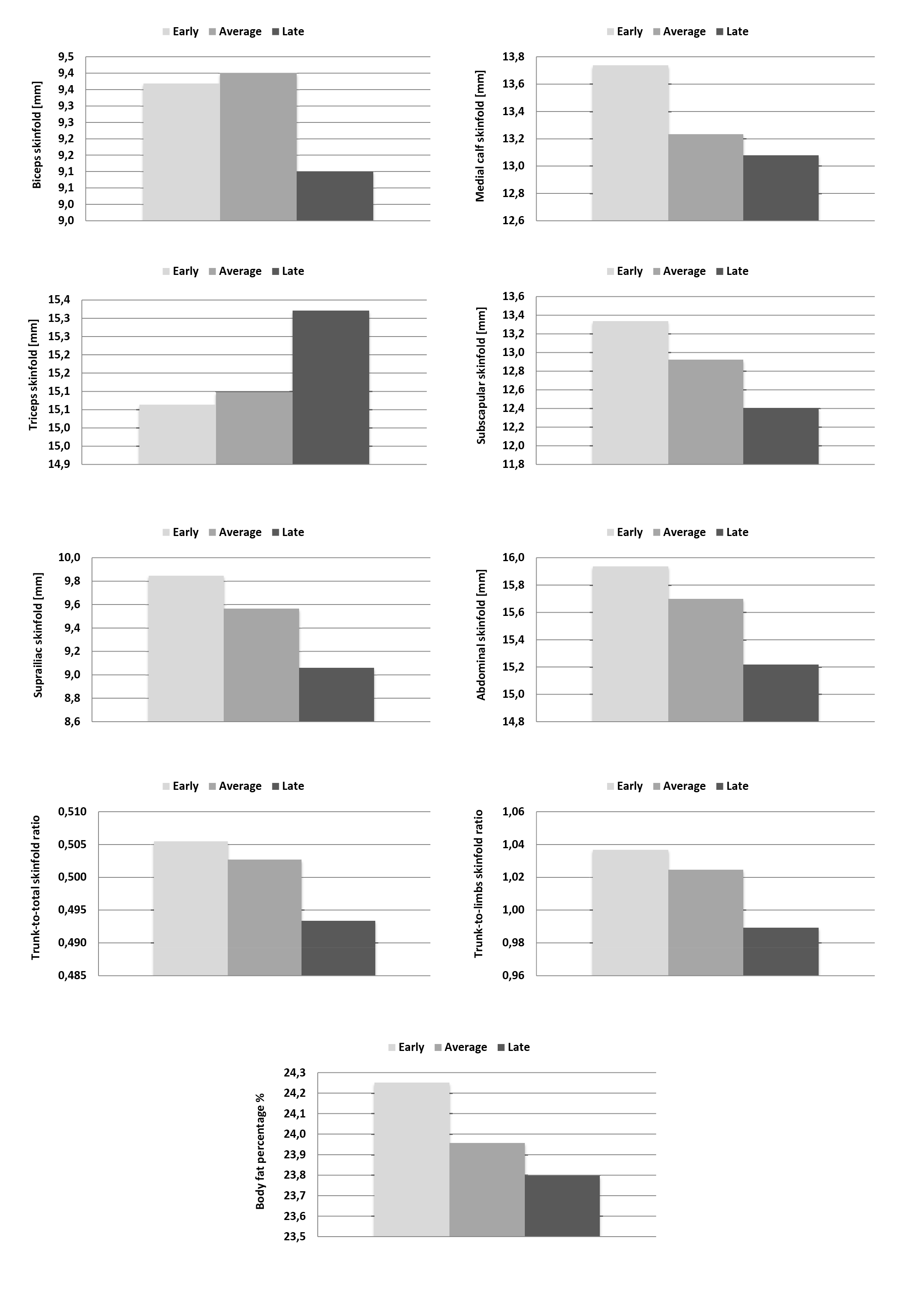
Early maturing girls were characterized by greater biceps and calf skinfolds compared to
their counterparts who achieved menarche late or at average age (Table 2). Interestingly, the triceps skinfold was, on average, the
thickest in late maturing girls. Yet, all differences in limbs adiposity were
statistically not significant (Table 2-5).
The highest thickness of subscapular, suprailiac, and abdominal skinfolds was noted in
the early maturing girls, while those experiencing menarche late and on average time had
lower values. Yet, the differences did not achieve statistical significance (Table 2, 4).
Table 2 Mean (X) and standard deviations (SD) values of various skinfold thicknesses of
Bengali girls (aged 7-21 years) in three menarche categories: early: < -1 SD,
average:X ± 1 SD, late: > 1 SD.
Menarcheal status
Triceps skinfold [mm]
Biceps skinfold [mm]
Subscapularskinfold [mm]
Suprailiac skinfold [mm]
Medialcalfskinfold [mm]
Abdominal skinfold [mm]
X
SD
X
SD
X
SD
x
SD
x
SD
x
SD
Early
15.1
4.1
9.4
3.7
13.3
4.0
9.8
3.5
13.7
4.2
15.9
4.8
Average
15.1
4.0
9.4
3.7
12.9
4.1
9.6
3.5
13.2
3.9
15.7
4.7
Late
15.3
4.0
9.1
3.6
12.4
3.8
9.1
3.5
13.1
3.8
15.2
5.0
Table 3 Mean (X) and standard deviations (SD) values of various skinfold ratios and body
fat of Bengali girls (aged 7-21 years) in three menarche categories: early:
< -1 SD, average: X ± 1 SD, late: > 1 SD.
Menarcheal status
Trunk-to-total skinfold ratio
Trunk-to-limbs skinfold ratio
Abdominal-to-trunk skinfold ratio
Body fat [%]
X
SD
X
SD
X
SD
X
SD
Early
0.506
0.042
1.037
0.177
0.408
0.041
24.3
5.1
Average
0.503
0.041
1.025
0.169
0.413
0.047
24.0
5.1
Late
0.493
0.044
0.989
0.182
0.416
0.042
23.8
4.9
Table 4 Difference between the mean values of various skinfold thicknesses of Bengali
girls (aged 7-21 years) in three menarche categories: early: < -1 SD, average:
X ± 1 SD, late: > 1 SD.
Comparison
Triceps skinfold [mm]
Biceps skinfold [mm]
Subscapular skinfold [mm]
Suprailiac skinfold [mm]
Medial calf skinfold [mm]
Abdominal skinfold [mm]
Average vs early
0.034
0.031
-0.414
-0.281
-0.503
-0.240
Average vs late
-0.223
0.299
0.515
0.506
0.155
0.478
Early vs late
-0.257
0.268
0.929
0.788
0.658
0.718
Table 5 Difference between the mean values of various skinfold ratios and body fat of
Bengali girls (aged 7-21 years) in three menarche categories: early: < -1 SD,
average: X ± 1 SD, late: > 1 SD.
Comparison
Trunk-to-total skinfold ratio
Trunk-to-limbs skinfold ratio
Abdominal-to-trunk skinfold ratio
Body adiposity [%]
Average vs early
-0.003
-0.012
0.005
-0.293
Average vs late
0.009***
0.035*
-0.003
0.161
Early vs late
0.012*
0.048*
-0.008
0.454
*p<0.05; ***p<0.0001
Girls and women with early and average-timed onset of menarche were also characterized by
lower values of trunk-to-total skinfold ratio, compared to their counterparts categorized
as having the first menstruation early. These differences mirror the relatively higher
trunk adiposity in early maturing girls. Noted discrepancies turned out to be
statistically significant between average and early, as well as late and early maturing
girls (Table 5).
This observation was confirmed by the highest value of trunk-to-limbs skinfold ratio in
early maturing girls due to greater accumulation of fat tissue on the trunk relative to
the limbs. The differences were statistically significant between average and early, as
well as late and early maturing girls (Table
5).
Early maturing individuals were characterized by greater general body fat. Yet, the
differences were not statistically significant (Table 3, 5).
Discussion
Age at menarche and overall adiposity
In the present study, early maturing girls were characterized by greater general body fat
ratio, in comparison to their counterparts who experienced menarche late or on an average
time. This is in line with a study conducted by Wilson et al. (Wilson et al. 2015), where younger menarcheal age was associated with
increased adiposity in young adult women. The findings are analogous to other large-scale
studies which have also shown an evident relationship between an early first menstruation
and greater adiposity in adulthood (Freedman et al.
2003; Garn et al. 1986; Trikudanathan et al. 2013).The present findings are
in line with a recent randomized study by Bell et al. (Bell et al. 2018) who noted that a relatively late first menstruation was
associated with lower adiposity later on. Increased obesity associated with relatively
early menarche appears to persist at least into middle adulthood (Dreyfus et al. 2015). Other studies noted that the tendency towards
greater overall adiposity tends to be observed in adolescence (Biro et al. 2003; Freedman et al.
2002).
The mechanism guiding the relationship between menarche and body fat ratio is still not
fully known. It was suggested, that both phenomena – adiposity and early maturation – are
not necessarily interdependent, but rather related to the same factor, namely excess
adiposity during childhood (Ong et al. 2007;
Lenthe et al. 1996), especially as it has
recently been shown that the relation between age at menarche and adiposity is largely
accounted for by body fat at the age of 8 (Bell et al.
2018). This further suggests that adiposity in childhood tends to track forward
to later life stages. Similar findings have been presented in previous studies (Johnson et al. 2015; Power et al. 1997). In this context, it should also be mentioned that
the described association may be caused by the differing pace of the maturation process,
that tends to be accelerated in individuals with relatively early puberty, in comparison
to late maturation. Consequently, the impairment of not only biological but also
behavioral processes that results from more rapid maturation alone can be associated with
an increased body fat ratio (Must et al. 2005;
Reynolds and Sontag 1950).
It has also been suggested that genetic factors associated with adiposity such as leptin
or insulin-like growth factor I (IGF-I) may play a role in the regulation of the onset of
puberty (Wilson et al. 2015).
It should be noted, that the differences in adiposity between early and late maturing
girls may be associated with different lifestyle choices, particularly in terms of
physical activity. For example, the authors of the CARDIA study reported higher levels of
physical activity in later-maturing girls, which, on top of influencing the body fat
ratio, may have delayed menarche (Chavarro et al.
2005).
It should be emphasized that elevated body adiposity related to early menarche can have
significant health-related consequences. Girls experiencing menarche at a relatively young
age are characterized by higher blood pressure in adulthood (Werneck et al. 2018). Additionally, it has also been suggested, that
a later (but within the normal range) pubertal onset is potentially protective against
obesity and cardio-metabolic risk in later life (Dreyfus
et al. 2015).
Age at menarche and body fat distribution
In the present study, early-maturing girls were characterized by a greater central
(android) adiposity, in comparison to their average and late maturing counterparts. We
consider surface anthropometric measurements to be able to accurately predict overall and
regional adiposity as measured by DXA method. Trunk adiposity is strongly correlated with
skinfold thickness, particularly suprailiac and abdominal skinfolds (around 90% and 60% of
the explained variability, respectively, for boys and girls) (Flavel et al. 2012; Leppik et al.
2004).
This is in line with a study in Portuguese girls. Higher waist circumferences were noted
in girls with earlier menarche. Differences in the body fat distribution occurred
regardless of differences in the body fat ratio (Leitão
et al. 2013). The present findings are analogous to the BioCycle study that
showed that early menarche was associated with a tendency towards central obesity (i.e.,
elevated trunk adiposity) (Chen et al. 2011).
Central adiposity has been linked to decreased insulin sensitivity, suggesting that early
menarche can be a predisposition to illnesses such as type II diabetes, hypertension,
coronary heart disease, stroke or metabolic syndrome. This was confirmed by a study
directly examining the relationship between the age of the first menstruation and insulin
resistance. Wilson et al. (Wilson et al. 2015)
found a 41% difference in insulin sensitivity between late and early maturing girls which
is consistent with studies linking younger menarcheal age with higher fasting insulin
concentration as well as increased insulin resistance, measured by homeostatic model
assessment for insulin resistance (HOMA-IR) (Chen et al.
2011; Feng et al. 2008). Dreyfus et al.
(Dreyfus et al. 2015) demonstrated that each
1-year of earlier maturation increased the risk of impaired fasting glycaemia (IFG) by 13%
and of metabolic syndrome by 19%. The risks increased independently of the overall body
fat ratio. This association has also been noted in Bangladeshi girls in whom the age at
menarche was inversely associated with the occurrence of the metabolic syndrome. An
analogous relationship was also shown in the presence of certain components (i.e., high
triglyceride, low HDL cholesterol) of the metabolic syndrome when examined individually
(Akter et al., 2012). The association of early menarche with an increased risk of
metabolic abnormalities was noted in various populations (Akter et al. 2012; Rah et al. 2009;
Stöckl et al. 2011). We concluded that these
phenomena are present regardless of ethnicity and the overall socioeconomic or
developmental status of a particular society.
Body composition during childhood, particularly at the age of 8 years, is critical to the
association between age at menarche and adiposity. On the other hand, the age of 12 has
been proposed as a point when obesity risks decline. This observation was made using
indicators of general and central adiposity (i.e., the waist circumference). This appears
to be consistent with other data that classify menarche under 12 years of age as early
maturation (Dreyfus et al. 2015; Kvalheim et al. 2016; Leitão et al. 2013; Werneck et al.
2018). In the analyzed population, the average age at menarche was 11.67 years,
with a standard deviation of 1.32. Hence, the cut-off point of early menarche was 10.36
years of age, which is lower than the previously mentioned threshold of the age of 12.
This difference may be associated with the specific population, i.e., middle-class West
Bengali girls. Most other research was conducted primarily in populations of Caucasian
origin.
The association between insulin resistance, glucose deregulation, central obesity, and
age at menarche is favoured by the metabolic and hormonal changes (i.e., modifications in
oestrogen, progesterone and sex hormone-binding globulin levels) in girls who mature at
earlier age, during a window of susceptibility (Prentice
and Viner 2013). Interestingly, in a large scale genetic study, there were no
significant associations of individual, central adiposity loci with the onset of menarche.
However, the authors pointed out, that their results may suggest a more complex genetic
relationship between the moment of the first menstruation and an android fat deposition
(Fernández-Rhodes et al. 2013).
Strengths and limitations
Among the strengths of the current study is the large number of participants from the same
population. In addition, the study was conducted in a city whose characteristics allow
extrapolation of the results to the entire Bengali population. The present study also had
some limitations. One of them is the use of the retrospective recall method to determine age
at menarche. According to some authors, this method may not be accurate enough because of
the time lag between menarche and the date of contact.
Conclusions, possible practical application and further research
Early, average and late maturing girls from the West Bengali population differed in terms
of body composition. The differences were particularly evident when body fat distribution
was taken into consideration, as individuals experiencing menarche relatively earlier were
characterized by greater trunk/abdominal adiposity in comparison to their average and late
maturing counterparts. We concluded that in this population, early menarche is associated
with a tendency towards a central (android) fat tissue distribution and, therefore, an
increased risk of abdominal obesity.
The present research, as well as a plethora of previous studies, showed that the tendency
towards elevated overall adiposity and central deposition of fat tissue in early maturing
girls persists into young adulthood. Furthermore, there is evidence that it may even be
present into middle age (Freedman et al. 2003;
Trikudanathan et al. 2013).
Given the association between body composition, fat distribution, and age at menarche, we
concluded that early maturation may predispose to abnormalities such as type II diabetes,
hypertension, coronary heart disease, stroke, or metabolic syndrome as these features have
been shown to be elevated in individuals with increased android deposition of adipose tissue
(Wilson et al. 2015).
Data on menarche may be critical in identifying women at risk for metabolic abnormalities
later in life. Early detection of such problems will, in turn, help prevent the metabolic
syndrome and, subsequently, also various cardiovascular diseases.
It should be noted that understanding the potential impact of menarche on a woman's future
metabolic health can be an important prophylactic tool to identify increased risk and
prevent the occurrence of metabolic abnormalities. This can often be achieved through simple
lifestyle and dietary habits, as these mediators are especially critical in younger
women.
Future research should investigate the relationship between age at menarche and metabolic
characteristics, preferably in ethnically diverse populations. It should also be emphasized
that longitudinal studies, as well as well-designed research conducted in large cohorts,
will be particularly valuable. In addition, it would be beneficial to adjust the results for
various lifestyle factors such as diet or physical activity, as well as for ethnically
determined characteristics related to body composition.
Appendix
Figure 1 Differences in mean values of analyzed characteristics between the girls in
three menarche categories.
Acknowledgements
This study has been sponsored by the Neys van Hoogstraten Foundation, The Netherlands
(ID158) and Indian Statistical Institute, Kolkata.
References
Akter, S./Jesmin, S./Islam, M./Sultana, S.
N./Okazaki, O./Hiroe, M./Moroi, M./Mizutani, T. (2012). Association of age at menarche
with metabolic syndrome and its components in rural Bangladeshi women. Nutrition &
Metabolism 9 (1), 99. https://doi.org/10.1186/1743-7075-9-99.
Al-Awadhi, N./Al-Kandari, N./Al-Hasan,
T./Almurjan, D./Ali, S./Al-Taiar, A. (2013). Age at menarche and its relationship to
body mass index among adolescent girls in Kuwait. BMC Public Health 13, 29. https://doi.org/10.1186/1471-2458-13-29.
Bagga, A./Kulkarni, S. (2000). Age at menarche and
secular trend in Maharashtrian (Indian) girls. Acta Biologica Szegediensis 44, 53–57.
Available online at http://www.sci.u-szeged.hu/ABS.
Batubara, J. R. L./Soesanti, F./Waal, H. D. van de
(2010). Age at menarche in Indonesian girls: a national survey. Acta Medica Indonesiana
42 (2), 78–81.
Bell, J. A./Carslake, D./Wade, K. H./Richmond, R.
C./Langdon, R. J./Vincent, E. E./Holmes, M. V./Timpson, N. J./Davey Smith, G. (2018).
Influence of puberty timing on adiposity and cardiometabolic traits: A Mendelian
randomisation study. PLoS Medicine 15 (8), e1002641. https://doi.org/10.1371/journal.pmed.1002641.
Biro, F. M./Lucky, A. W./Simbartl, L. A./Barton,
B. A./Daniels, S. R./Striegel-Moore, R./Kronsberg, S. S./Morrison, J. A. (2003).
Pubertal maturation in girls and the relationship to anthropometric changes: pathways
through puberty. The Journal of Pediatrics 142 (6), 643–646. https://doi.org/10.1067/mpd.2003.244.
Chang, S. H./Tzeng, S. J./Cheng, J. Y./Chie, W. C.
(2000). Height and weight change across menarche of schoolgirls with early menarche.
Archives of Pediatrics & Adolescent Medicine 154 (9), 880–884. https://doi.org/10.1001/archpedi.154.9.880.
Chavarro, J. E./Peterson, K. E./Sobol, A.
M./Wiecha, J. L./Gortmaker, S. L. (2005). Effects of a school-based obesity-prevention
intervention on menarche (United States). Cancer Causes & Control : CCC 16 (10),
1245–1252. https://doi.org/10.1007/s10552-005-0404-5.
Chen, L./Zhang, C./Yeung, E./Ye, A./Mumford, S.
L./Wactawski-Wende, J./Schisterman, E. F. (2011). Age at menarche and metabolic markers
for type 2 diabetes in premenopausal women: the BioCycle Study. The Journal of Clinical
Endocrinology and Metabolism 96 (6), E1007-12. https://doi.org/10.1210/jc.2010-2526.
Deardorff, J./Abrams, B./Ekwaru, J. P./Rehkopf, D.
H. (2014). Socioeconomic status and age at menarche: an examination of multiple
indicators in an ethnically diverse cohort. Annals of Epidemiology 24 (10), 727–733.
https://doi.org/10.1016/J.ANNEPIDEM.2014.07.002.
Demerath, E. W./Liu, C.-T./Franceschini, N./Chen,
G./Palmer, J. R./Smith, E. N./Chen, C. T. L./Ambrosone, C. B./Arnold, A. M./Bandera, E.
V./Berenson, G. S./Bernstein, L./Britton, A./Cappola, A. R./Carlson, C. S./Chanock, S.
J./Chen, W./Chen, Z./Deming, S. L./Elks, C. E./Evans, M. K./Gajdos, Z./Henderson, B.
E./Hu, J. J./Ingles, S./John, E. M./Kerr, K. F./Kolonel, L. N./Le Marchand, L./Lu,
X./Millikan, R. C./Musani, S. K./Nock, N. L./North, K./Nyante, S./Press, M.
F./Rodriquez-Gil, J. L./Ruiz-Narvaez, E. A./Schork, N. J./Srinivasan, S. R./Woods, N.
F./Zheng, W./Ziegler, R. G./Zonderman, A./Heiss, G./Gwen Windham, B./Wellons, M./Murray,
S. S./Nalls, M./Pastinen, T./Rajkovic, A./Hirschhorn, J./Adrienne Cupples,
L./Kooperberg, C./Murabito, J. M./Haiman, C. A. (2013). Genome-wide association study of
age at menarche in African-American women. Human Molecular Genetics 22 (16), 3329–3346.
https://doi.org/10.1093/hmg/ddt181.
Dreyfus, J./Jacobs, D. R./Mueller, N./Schreiner,
P. J./Moran, A./Carnethon, M. R./Demerath, E. W. (2015). Age at menarche and
cardiometabolic risk in adulthood: The coronary artery risk development in young adults
study. The Journal of Pediatrics 167 (2), 344-52.e1. https://doi.org/10.1016/j.jpeds.2015.04.032.
Elks, C. E./Perry, J. R. B./Sulem, P./Chasman, D.
I./Franceschini, N./He, C./Lunetta, K. L./Visser, J. A./Byrne, E. M./Cousminer, D.
L./Gudbjartsson, D. F./Esko, T./Feenstra, B./Hottenga, J.-J./Koller, D. L./Kutalik,
Z./Lin, P./Mangino, M./Marongiu, M./McArdle, P. F./Smith, A. V./Stolk, L./van Wingerden,
S. H./Zhao, J. H./Albrecht, E./Corre, T./Ingelsson, E./Hayward, C./Magnusson, P. K.
E./Smith, E. N./Ulivi, S./Warrington, N. M./Zgaga, L./Alavere, H./Amin, N./Aspelund,
T./Bandinelli, S./Barroso, I./Berenson, G. S./Bergmann, S./Blackburn, H./Boerwinkle,
E./Buring, J. E./Busonero, F./Campbell, H./Chanock, S. J./Chen, W./Cornelis, M.
C./Couper, D./Coviello, A. D./d'Adamo, P./Faire, U. de/Geus, E. J. C. de/Deloukas,
P./Döring, A./Smith, G. D./Easton, D. F./Eiriksdottir, G./Emilsson, V./Eriksson,
J./Ferrucci, L./Folsom, A. R./Foroud, T./Garcia, M./Gasparini, P./Geller, F./Gieger,
C./Gudnason, V./Hall, P./Hankinson, S. E./Ferreli, L./Heath, A. C./Hernandez, D.
G./Hofman, A./Hu, F. B./Illig, T./Järvelin, M.-R./Johnson, A. D./Karasik, D./Khaw,
K.-T./Kiel, D. P./Kilpeläinen, T. O./Kolcic, I./Kraft, P./Launer, L. J./Laven, J. S.
E./Li, S./Liu, J./Levy, D./Martin, N. G./McArdle, W. L./Melbye, M./Mooser, V./Murray, J.
C./Murray, S. S./Nalls, M. A./Navarro, P./Nelis, M./Ness, A. R./Northstone, K./Oostra,
B. A./Peacock, M./Palmer, L. J./Palotie, A./Paré, G./Parker, A. N./Pedersen, N.
L./Peltonen, L./Pennell, C. E./Pharoah, P./Polasek, O./Plump, A. S./Pouta, A./Porcu,
E./Rafnar, T./Rice, J. P./Ring, S. M./Rivadeneira, F./Rudan, I./Sala, C./Salomaa,
V./Sanna, S./Schlessinger, D./Schork, N. J./Scuteri, A./Segrè, A. V./Shuldiner, A.
R./Soranzo, N./Sovio, U./Srinivasan, S. R./Strachan, D. P./Tammesoo, M.-L./Tikkanen,
E./Toniolo, D./Tsui, K./Tryggvadottir, L./Tyrer, J./Uda, M./Dam, R. M. van/Meurs, J. B.
J. van/Vollenweider, P./Waeber, G./Wareham, N. J./Waterworth, D. M./Weedon, M.
N./Wichmann, H. E./Willemsen, G./Wilson, J. F./Wright, A. F./Young, L./Zhai, G./Zhuang,
W. V./Bierut, L. J./Boomsma, D. I./Boyd, H. A./Crisponi, L./Demerath, E. W./Duijn, C. M.
van/Econs, M. J./Harris, T. B./Hunter, D. J./Loos, R. J. F./Metspalu, A./Montgomery, G.
W./Ridker, P. M./Spector, T. D./Streeten, E. A./Stefansson, K./Thorsteinsdottir,
U./Uitterlinden, A. G./Widen, E./Murabito, J. M./Ong, K. K./Murray, A. (2010). Thirty
new loci for age at menarche identified by a meta-analysis of genome-wide association
studies. Nature Genetics 42 (12), 1077–1085. https://doi.org/10.1038/ng.714.
Feng, Y./Hong, X./Wilker, E./Li, Z./Zhang, W./Jin,
D./Liu, X./Zang, T./Xu, X./Xu, X. (2008). Effects of age at menarche, reproductive
years, and menopause on metabolic risk factors for cardiovascular diseases.
Atherosclerosis 196 (2), 590–597. https://doi.org/10.1016/j.atherosclerosis.2007.06.016.
Fernández-Rhodes, L./Demerath, E. W./Cousminer, D.
L./Tao, R./Dreyfus, J. G./Esko, T./Smith, A. V./Gudnason, V./Harris, T. B./Launer,
L./McArdle, P. F./Yerges-Armstrong, L. M./Elks, C. E./Strachan, D. P./Kutalik,
Z./Vollenweider, P./Feenstra, B./Boyd, H. A./Metspalu, A./Mihailov, E./Broer,
L./Zillikens, M. C./Oostra, B./Duijn, C. M. van/Lunetta, K. L./Perry, J. R. B./Murray,
A./Koller, D. L./Lai, D./Corre, T./Toniolo, D./Albrecht, E./Stöckl, D./Grallert,
H./Gieger, C./Hayward, C./Polasek, O./Rudan, I./Wilson, J. F./He, C./Kraft, P./Hu, F.
B./Hunter, D. J./Hottenga, J.-J./Willemsen, G./Boomsma, D. I./Byrne, E. M./Martin, N.
G./Montgomery, G. W./Warrington, N. M./Pennell, C. E./Stolk, L./Visser, J. A./Hofman,
A./Uitterlinden, A. G./Rivadeneira, F./Lin, P./Fisher, S. L./Bierut, L. J./Crisponi,
L./Porcu, E./Mangino, M./Zhai, G./Spector, T. D./Buring, J. E./Rose, L. M./Ridker, P.
M./Poole, C./Hirschhorn, J. N./Murabito, J. M./Chasman, D. I./Widen, E./North, K.
E./Ong, K. K./Franceschini, N. (2013). Association of adiposity genetic variants with
menarche timing in 92,105 women of European descent. American Journal of Epidemiology
178 (3), 451–460. https://doi.org/10.1093/aje/kws473.
Flavel, N. A./Olds, T. S./Buckley, J. D./Haren, M.
T./Petkov, J. (2012). Anthropometric estimates of total and regional body fat in
children aged 6-17 years. Acta Paediatrica : nurturing the Child 101 (12), 1253–1259.
https://doi.org/10.1111/APA.12024.
Freedman, D. S./Khan, L. K./Serdula, M. K./Dietz,
W. H./Srinivasan, S. R./Berenson, G. S. (2002). Relation of age at menarche to race,
time period, and anthropometric dimensions: the Bogalusa Heart Study. Pediatrics 110
(4), e43. https://doi.org/10.1542/PEDS.110.4.E43.
Freedman, D. S./Khan, L. K./Serdula, M. K./Dietz,
W. H./Srinivasan, S. R./Berenson, G. S. (2003). The relation of menarcheal age to
obesity in childhood and adulthood: the Bogalusa heart study. BMC Pediatrics 3, 3.
https://doi.org/10.1186/1471-2431-3-3.
Gajdos, Z. K. Z./Henderson, K. D./Hirschhorn, J.
N./Palmert, M. R. (2010). Genetic determinants of pubertal timing in the general
population. Molecular and Cellular Endocrinology 324 (1-2), 21–29. https://doi.org/10.1016/j.mce.2010.01.038.
Garn, S. M./LaVelle, M./Rosenberg, K.
R./Hawthorne, V. M. (1986). Maturational timing as a factor in female fatness and
obesity. The American Journal of Clinical Nutrition 43 (6), 879–883. https://doi.org/10.1093/ajcn/43.6.879.
Gomula, A./Koziel, S. (2018). Secular trend and
social variation in age at menarche among Polish schoolgirls before and after the
political transformation. American Journal of Human Biology 30 (1). https://doi.org/10.1002/ajhb.23048.
Gupta, A. K./Vatsayan, A./Ahluwalia, S. K./Sood,
R. K./Mazta, S. R./Sharma, R. (1996). Age at menarche, menstrual knowledge and practices
in the apple belt of Shimla hills. Journal of Obstetrics and Gynaecology 16 (6),
548–550. https://doi.org/10.3109/01443619609030096.
Harris, M. A./Prior, J. C./Koehoorn, M. (2008).
Age at menarche in the Canadian population: secular trends and relationship to adulthood
BMI. The Journal of Adolescent Health 43 (6), 548–554. https://doi.org/10.1016/J.JADOHEALTH.2008.07.017.
Hermanussen, M./Lehmann, A./Scheffler, C. (2012).
Psychosocial pressure and menarche: a review of historic evidence for social amenorrhea.
Obstetrical & Gynecological Survey 67 (4), 237–241. https://doi.org/10.1097/OGX.0B013E31824C94AD.
James-Todd, T./Tehranifar, P./Rich-Edwards,
J./Titievsky, L./Terry, M. B. (2010). The impact of socioeconomic status across early
life on age at menarche among a racially diverse population of girls. Annals of
Epidemiology 20 (11), 836–842. https://doi.org/10.1016/J.ANNEPIDEM.2010.08.006.
John, J./Verma, M./Chhatwal, J. (2014).
Physiological variables, psychosocial factors and age at menarche among Punjabi girls.
Indian Journal of Physiology and Pharmacology 58 (2), 141–146.
Johnson, W./Li, L./Kuh, D./Hardy, R. (2015). How
has the age-related process of overweight or obesity development changed over time?
Co-ordinated analyses of individual participant data from five United Kingdom birth
cohorts. PLoS Medicine 12 (5), e1001828; discussion e1001828. https://doi.org/10.1371/journal.pmed.1001828.
Kozieł, S./Gomula, A./Nowak-Szczepańska, N.
(2016). The association between social factors and body length proportions in Polish
schoolchildren from Lower Silesia. Anthropological Review 79 (4), 397–408. https://doi.org/10.1515/anre-2016-0029.
Kvalheim, S./Sandvik, L./Winsvold, B./Hagen,
K./Zwart, J.-A. (2016). Early menarche and chronic widespread musculoskeletal
complaints--Results from the HUNT study. European Journal of Pain 20 (3), 458–464.
https://doi.org/10.1002/ejp.747.
Lakshman, R./Forouhi, N./Luben, R./Bingham,
S./Khaw, K./Wareham, N./Ong, K. K. (2008). Association between age at menarche and risk
of diabetes in adults: results from the EPIC-Norfolk cohort study. Diabetologia 51 (5),
781–786. https://doi.org/10.1007/s00125-008-0948-5.
Leitão, R. B./Rodrigues, L. P./Neves, L./Carvalho,
G. S. (2013). Development of adiposity, obesity and age at menarche: an 8-year follow-up
study in Portuguese schoolgirls. International Journal of Adolescent Medicine and Health
25 (1), 55–63. https://doi.org/10.1515/ijamh-2013-0007.
Lenthe, F. J. van/Kemper, C. G./Mechelen, W. van
(1996). Rapid maturation in adolescence results in greater obesity in adulthood: the
Amsterdam Growth and Health Study. The American Journal of Clinical Nutrition 64 (1),
18–24. https://doi.org/10.1093/ajcn/64.1.18.
Leppik, A./Jürimäe, T./Jürimäe, J. (2004).
Influence of anthropometric parameters on the body composition measured by bioelectrical
impedance analysis or DXA in children. Acta Paediatrica : nurturing the Child 93 (8),
1036–1041. https://doi.org/10.1111/J.1651-2227.2004.TB02714.X.
Mueller, N. T./Duncan, B. B./Barreto, S. M./Chor,
D./Bessel, M./Aquino, E. M. L./Pereira, M. A./Schmidt, M. I. (2014). Earlier age at
menarche is associated with higher diabetes risk and cardiometabolic disease risk
factors in Brazilian adults: Brazilian Longitudinal Study of Adult Health (ELSA-Brasil).
Cardiovascular Diabetology 13, 22. https://doi.org/10.1186/1475-2840-13-22.
Mumm, R./Scheffler, C./Hermanussen, M. (2014).
Developing differential height, weight and body mass index references for girls that
reflect the impact of the menarche. Acta Paediatrica: nurturing the Child 103 (7),
e312-6. https://doi.org/10.1111/apa.12625.
Must, A./Naumova, E. N./Phillips, S. M./Blum,
M./Dawson-Hughes, B./Rand, W. M. (2005). Childhood overweight and maturational timing in
the development of adult overweight and fatness: the Newton Girls Study and its
follow-up. Pediatrics 116 (3), 620–627. https://doi.org/10.1542/peds.2004-1604.
Ong, K. K./Northstone, K./Wells, J. C. K./Rubin,
C./Ness, A. R./Golding, J./Dunger, D. B. (2007). Earlier mother's age at menarche
predicts rapid infancy growth and childhood obesity. PLoS Medicine 4 (4), e132.
https://doi.org/10.1371/journal.pmed.0040132.
Onland-Moret, N. C./Peeters, P. H. M./van Gils, C.
H./Clavel-Chapelon, F./Key, T./Tjønneland, A./Trichopoulou, A./Kaaks, R./Manjer,
J./Panico, S./Palli, D./Tehard, B./Stoikidou, M./Bueno-De-Mesquita, H. B./Boeing,
H./Overvad, K./Lenner, P./Quirós, J. R./Chirlaque, M. D./Miller, A. B./Khaw, K.
T./Riboli, E. (2005). Age at menarche in relation to adult height: the EPIC study.
American Journal of Epidemiology 162 (7), 623–632. https://doi.org/10.1093/aje/kwi260.
Perry, J. R./Day, F./Elks, C. E./Sulem,
P./Thompson, D. J./Ferreira, T./He, C./Chasman, D. I./Esko, T./Thorleifsson,
G./Albrecht, E./Ang, W. Q./Corre, T./Cousminer, D. L./Feenstra, B./Franceschini,
N./Ganna, A./Johnson, A. D./Kjellqvist, S./Lunetta, K. L./McMahon, G./Nolte, I.
M./Paternoster, L./Porcu, E./Smith, A. V./Stolk, L./Teumer, A./Tšernikova, N./Tikkanen,
E./Ulivi, S./Wagner, E. K./Amin, N./Bierut, L. J./Byrne, E. M./Hottenga, J.-J./Koller,
D. L./Mangino, M./Pers, T. H./Yerges-Armstrong, L. M./Zhao, J. H./Andrulis, I.
L./Anton-Culver, H./Atsma, F./Bandinelli, S./Beckmann, M. W./Benitez, J./Blomqvist,
C./Bojesen, S. E./Bolla, M. K./Bonanni, B./Brauch, H./Brenner, H./Buring, J.
E./Chang-Claude, J./Chanock, S./Chen, J./Chenevix-Trench, G./Collée, J. M./Couch, F.
J./Couper, D./Coveillo, A. D./Cox, A./Czene, K./D'adamo, A. P./Smith, G. D./Vivo, I.
de/Demerath, E. W./Dennis, J./Devilee, P./Dieffenbach, A. K./Dunning, A.
M./Eiriksdottir, G./Eriksson, J. G./Fasching, P. A./Ferrucci, L./Flesch-Janys,
D./Flyger, H./Foroud, T./Franke, L./Garcia, M. E./García-Closas, M./Geller, F./Geus, E.
E. de/Giles, G. G./Gudbjartsson, D. F./Gudnason, V./Guénel, P./Guo, S./Hall, P./Hamann,
U./Haring, R./Hartman, C. A./Heath, A. C./Hofman, A./Hooning, M. J./Hopper, J. L./Hu, F.
B./Hunter, D. J./Karasik, D./Kiel, D. P./Knight, J. A./Kosma, V.-M./Kutalik, Z./Lai,
S./Lambrechts, D./Lindblom, A./Mägi, R./Magnusson, P. K./Mannermaa, A./Martin, N.
G./Masson, G./McArdle, P. F./McArdle, W. L./Melbye, M./Michailidou, K./Mihailov,
E./Milani, L./Milne, R. L./Nevanlinna, H./Neven, P./Nohr, E. A./Oldehinkel, A.
J./Oostra, B. A./Palotie, A./Peacock, M./Pedersen, N. L./Peterlongo, P./Peto,
J./Pharoah, P. D./Postma, D. S./Pouta, A./Pylkäs, K./Radice, P./Ring, S./Rivadeneira,
F./Robino, A./Rose, L. M./Rudolph, A./Salomaa, V./Sanna, S./Schlessinger, D./Schmidt, M.
K./Southey, M. C./Sovio, U./Stampfer, M. J./Stöckl, D./Storniolo, A. M./Timpson, N.
J./Tyrer, J./Visser, J. A./Vollenweider, P./Völzke, H./Waeber, G./Waldenberger,
M./Wallaschofski, H./Wang, Q./Willemsen, G./Winqvist, R./Wolffenbuttel, B. H./Wright, M.
J./Boomsma, D. I./Econs, M. J./Khaw, K.-T./Loos, R. J./McCarthy, M. I./Montgomery, G.
W./Rice, J. P./Streeten, E. A./Thorsteinsdottir, U./van Duijn, C. M./Alizadeh, B.
Z./Bergmann, S./Boerwinkle, E./Boyd, H. A./Crisponi, L./Gasparini, P./Gieger, C./Harris,
T. B./Ingelsson, E./Järvelin, M.-R./Kraft, P./Lawlor, D./Metspalu, A./Pennell, C.
E./Ridker, P. M./Snieder, H./Sørensen, T. I./Spector, T. D./Strachan, D.
P./Uitterlinden, A. G./Wareham, N. J./Widen, E./Zygmunt, M./Murray, A./Easton, D.
F./Stefansson, K./Murabito, J. M./Ong, K. K. (2014). Parent-of-origin-specific allelic
associations among 106 genomic loci for age at menarche. Nature 514 (7520), 92–97.
https://doi.org/10.1038/nature13545.
Power, C./Lake, J. K./Cole, T. J. (1997). Body
mass index and height from childhood to adulthood in the 1958 British born cohort. The
American Journal of Clinical Nutrition 66 (5), 1094–1101. https://doi.org/10.1093/ajcn/66.5.1094.
Prentice, P./Viner, R. M. (2013). Pubertal timing
and adult obesity and cardiometabolic risk in women and men: a systematic review and
meta-analysis. International Journal of Obesity 37 (8), 1036–1043. https://doi.org/10.1038/IJO.2012.177.
Rah, J. H./Shamim, A. A./Arju, U. T./Labrique, A.
B./Rashid, M./Christian, P. (2009). Age of onset, nutritional determinants, and seasonal
variations in menarche in rural Bangladesh. Journal of Health, Population, and Nutrition
27 (6), 802–807. https://doi.org/10.3329/JHPN.V27I6.4332.
Reynolds, E. L./Sontag, L. W. (1950). The
distribution of subcutaneous fat in childhood and adolescence. Monographs of the Society
for Research in Child Development 15 (2), iii-189. https://doi.org/10.2307/1165600.
Slaughter, M. H./Lohman, T. G./Boileau, R.
A./Horswill, C. A./Stillman, R. J./van Loan, M. D./Bemben, D. A. (1988). Skinfold
equations for estimation of body fatness in children and youth. Human biology 60 (5),
709–723.
Stöckl, D./Meisinger, C./Peters, A./Thorand,
B./Huth, C./Heier, M./Rathmann, W./Kowall, B./Stöckl, H./Döring, A. (2011). Age at
menarche and its association with the metabolic syndrome and its components: results
from the KORA F4 study. PloS one 6 (10), e26076. https://doi.org/10.1371/journal.pone.0026076.
Tanner, J. M. (1962). Growth at adolescence. With
a general consideration of the effects of hereditary and environmental factors upon
growth and maturation from birth to maturity. 2nd ed. Oxford, Blackwell
Scientific.
Trikudanathan, S./Pedley, A./Massaro, J.
M./Hoffmann, U./Seely, E. W./Murabito, J. M./Fox, C. S. (2013). Association of female
reproductive factors with body composition: the Framingham Heart Study. The Journal of
Clinical Endocrinology and Metabolism 98 (1), 236–244. https://doi.org/10.1210/jc.2012-1785.
Weiner, J. S./Lourie, J. A. (1969). Human biology.
A guide to field methods. Oxford, Blackwell Scientific.
Werneck, A. O./Oyeyemi, A. L./Cyrino, E.
S./Ronque, E. R. V./Szwarcwald, C. L./Coelho-E-Silva, M. J./Silva, D. R. (2018).
Association between age at menarche and blood pressure in adulthood: is obesity an
important mediator? Hypertension research : official journal of the Japanese Society of
Hypertension 41 (10), 856–864. https://doi.org/10.1038/s41440-018-0079-4.
Wilson, D. A./Derraik, J. G. B./Rowe, D.
L./Hofman, P. L./Cutfield, W. S. (2015). Earlier menarche is associated with lower
insulin sensitivity and increased adiposity in young adult women. PloS one 10 (6),
e0128427. https://doi.org/10.1371/journal.pone.0128427.
Żegleń, M./Marini, E./Cabras, S./Kryst, Ł./Das,
R./Chakraborty, A./Dasgupta, P. (2020). The relationship among the age at menarche,
anthropometric characteristics, and socio-economic factors in Bengali girls from
Kolkata, India. American Journal of Human Biology 32 (4), e23380. https://doi.org/10.1002/ajhb.23380.
 ✉
✉